Microtubules In A Cell Diagram Biology Diagrams
Microtubules In A Cell Diagram Biology Diagrams Fig 2 - Summary diagram showing the stages of mitosis. Clinical Relevance - Errors of Mitosis. Errors in mitosis typically occur during metaphase. Usually, this is due to a misalignment of chromosomes along the metaphase plate or a failure of the mitotic spindles to attach to one of the kinetochores. This can result in the daughter cells Mitosis Stages Diagram. Mitosis is the process of cell division in which a cell duplicates its genetic material and divides into two daughter cells. This process is essential for the growth, development, and maintenance of multicellular organisms. The mitotic spindle is a structure made up of microtubules that help move the chromosomes
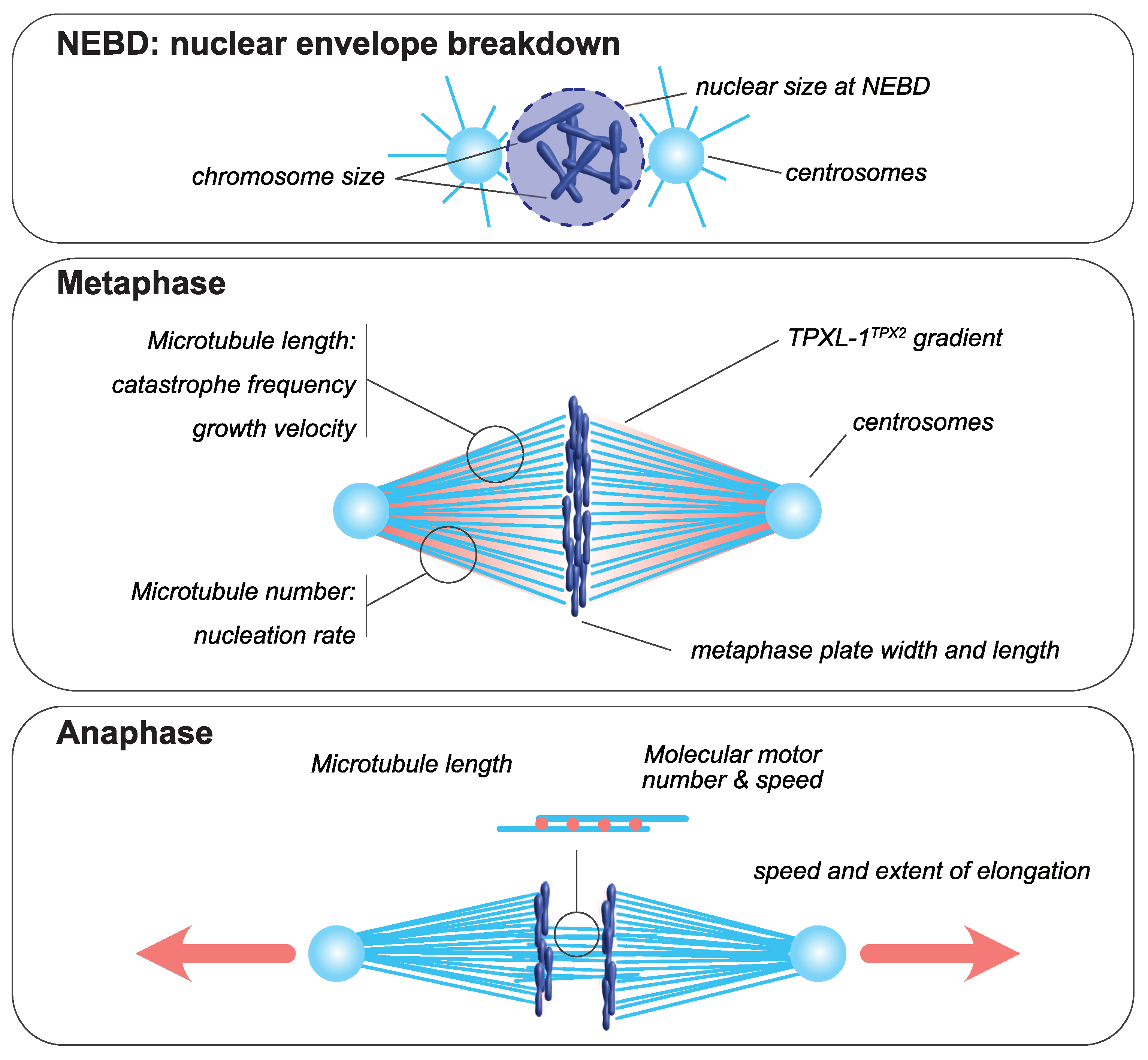
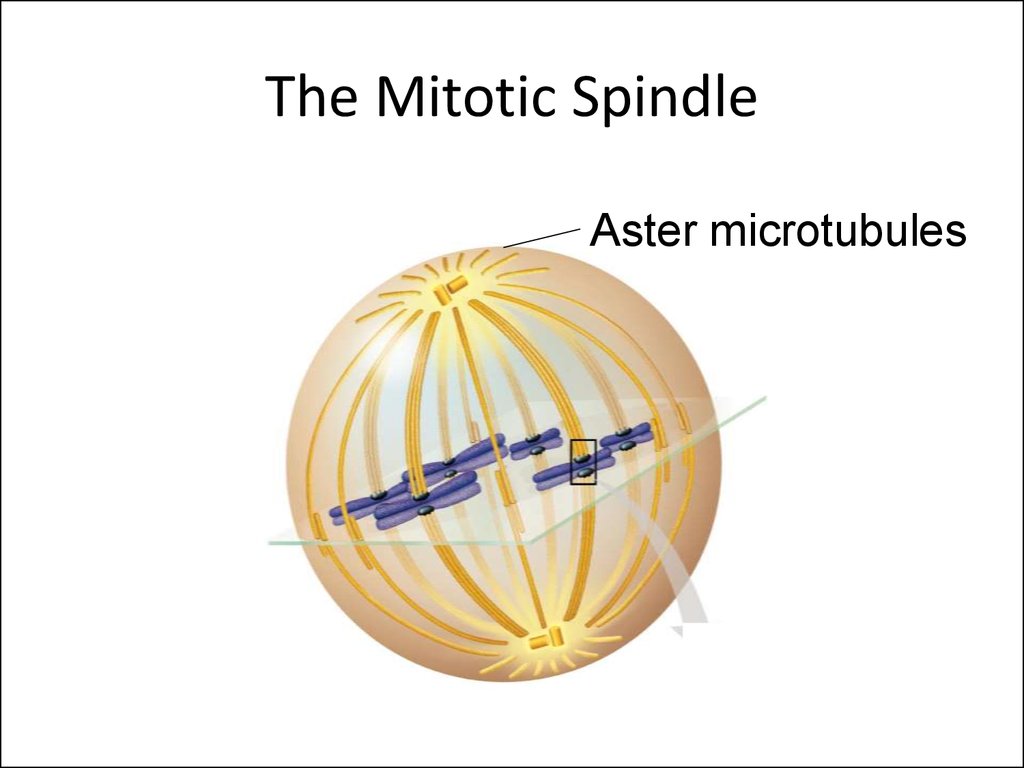
The nuclear envelope breaks down and spindles form at opposite poles of the cell. Prophase (versus interphase) is the first true step of the mitotic process. During prophase, several important changes occur: Chromatin fibers become coiled into chromosomes, with each chromosome having two chromatids joined at a centromere. This diagram depicts the organization of a typical mitotic spindle found in animal cells. Chromosomes are attached to kinetochore microtubules via a multiprotein complex called the kinetochore. Polar microtubules interdigitate at the spindle midzone and push the spindle poles apart via motor proteins. Astral microtubules anchor the spindle poles to the cell membrane. Learn about the stages and mechanisms of mitosis, the process of nuclear division in eukaryotic cells. See diagrams of the mitotic spindle, the structure that aids in chromosome separation.
The Stages of Mitosis and Cell Division Biology Diagrams
Explore the stages of mitosis with detailed diagrams. Understand each phase and discover real-world applications of this essential cell division process. The constriction invariably occurs in the plane of the metaphase plate, at right angles to the long axis of the mitotic spindle apparatus. The constriction grows more in-depth from the Download scientific diagram | Schematic showing a mitotic spindle inside a cell. (b) A simplified 1D force balance model. Two poles (r1 and r4) and one pair of sister chromatids (r2 and r3) are One effective way to understand the stages of mitosis is through a diagram with labels. This diagram provides a visual representation of the different phases and helps in comprehending the complex process. the nuclear envelope disintegrates, and the mitotic spindle begins to form. Prometaphase is marked by the fragmentation of the nuclear