Vasculature of the Heart Biology Diagrams
Vasculature of the Heart Biology Diagrams The coronary arteries are the arterial blood vessels of coronary circulation, which transport oxygenated blood to the heart muscle.The heart requires a continuous supply of oxygen to function and survive, much like any other tissue or organ of the body. [1]The coronary arteries wrap around the entire heart. The two main branches are the left coronary artery and right coronary artery.
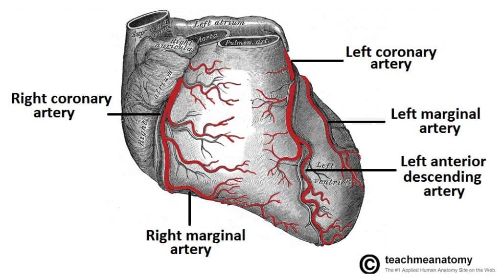
Gross anatomy. The typical configuration consists of two coronary arteries, a left main coronary artery (LMCA) and a right coronary artery (RCA), arising from the left (posterior) and right (anterior) aortic or coronary sinuses, respectively, in the proximal ascending aorta.These are the only two branches of the ascending aorta. The right coronary artery courses in the right atrioventricular The coronary arteries run along the coronary sulcus of the myocardium of the heart. Their main function is to supply blood to the heart. This is a crucial function for myocardial function and subsequently homeostasis of the body. The arrangement of coronary arteries varies among people significantly.

Coronary arteries and cardiac veins: Anatomy and branches Biology Diagrams
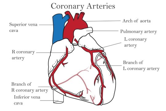
The coronary artery branches are the first of many branches off your aorta. Coronary artery structure. There are two coronary arteries, each containing several branches: Right coronary artery (RCA): The RCA supplies blood to your right atrium and right ventricle (where deoxygenated blood goes before heading to the lungs). Its branches supply

Smaller branches of the coronary arteries include: obtuse marginal (OM), septal perforator (SP), and diagonals. Why are the coronary arteries important? Since coronary arteries deliver blood to the heart muscle, any coronary artery disorder or disease can have serious implications by reducing the flow of oxygen and nutrients to the heart muscle.
Radiology Reference Article - Radiopaedia.org Biology Diagrams
Coronary arteries. The coronary arteries arise from the root of the ascending aorta.Recall that the aortic valve has three semilunar cusps, also known as the sinuses of Valsalva. The left and right semilunar cusps give rise to the corresponding left and right coronary arteries (respectively). The third sinus - which is the posterior semilunar cusp - is not associated with a coronary vessel
